
RUSH Protocol
Teaching graphics for ultrasound in shock
22 editable diagrams for your slides. Probe position, ultrasound views and findings in one style.
Buy the EN pack on Gumroad From CHF 5 · Suggested CHF 7
What’s in the pack
The pack includes editable SVGs, PowerPoint slides and PDFs.
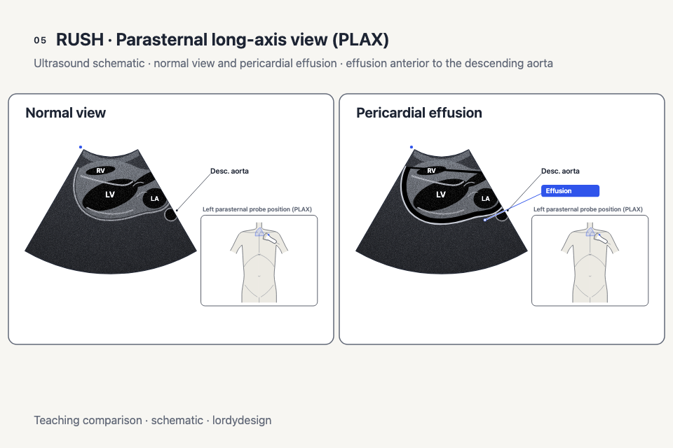
Pump
Cardiac probe positions, views of the heart, pericardial effusion and ventricular function.
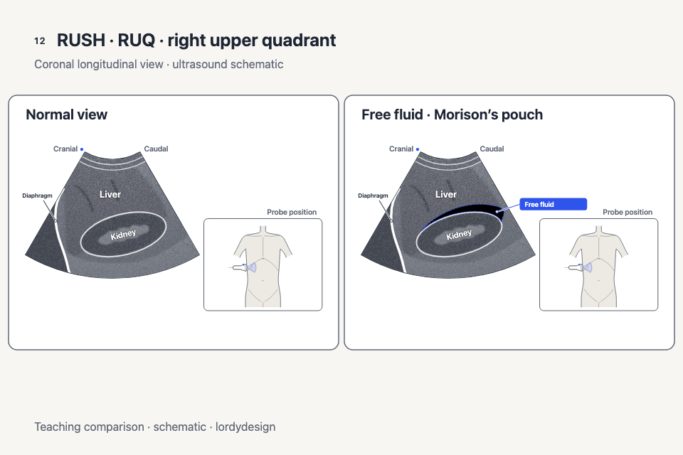
Tank
Inferior vena cava, abdominal and pelvic views, pleural findings and lung artifacts.
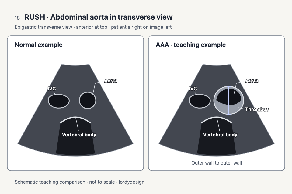
Pipes
Aortic views and diagrams of venous compression.


The pack also includes an overview of probe positions, a comparison of shock patterns and a card on common pitfalls.
Use in teaching
Introduce the views
Use the templates in a lecture or before a hands-on ultrasound session to connect probe positions with the corresponding views.
Compare diagrams
Place examples side by side and ask learners what changes and what stays the same.
Build a quiz
Hide labels in the editable SVGs for a short review exercise.
Teaching material only. The diagrams are not a medical device and do not replace clinical training or judgment.
Frequently asked questions
What file formats are included?
Editable SVGs, PowerPoint slides and PDF.
Are these real ultrasound images?
No. The pack uses schematic drawings.
Can I use the pack to make a diagnosis?
No. The illustrations are for teaching. The comparison of shock patterns is not exhaustive and does not rule out mixed patterns.